
Status: available
Supervisor: Michael Reiter, Christopher Pramerdorfer
The purpose of this work is to classify sequences of dermatoscopy images as “changing” or “not changing”. The term “change” refers to a depicted mole or skin lesion, and a given sequence should be classified as “changing” if the depicted mole changes in a form that indicates a tumor, not simply any change. Every sequence contains two or more images of the same mole. For a given sequence, the camera model might vary and the extrinsics are generally different but similar between images.
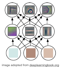 The envisioned approach for solving this problem is Deep Learning (DL) [1], the state of the art in many related problems including medical image analysis. A promising method is to perform image-level analysis using Convolutional Neural Networks (CNNs) [1] and to model temporal dependencies using graphical models (e.g. Bayesian networks) or LSTMs [2]. An alternative is to integrate these steps, for instance using 3D CNNs [3]. The main challenges in solving this problem are changes in the camera intrinsics and extrinsics between sequences and images in a given sequence, and the fact that not all changes in a mole equal “change” in the context of this problem. In order to overcome these challenges, corresponding preprocessing and/or data augmentation strategies will be designed.
Workflow
- Literature Review – getting to know the algorithms
- Data Preparation (database available)
- Design and Implementation of Deep Network Architekture
- Experiments and Evaluation
- Written Report/Thesis and final presentation
Requirements
- Experience with Matlab or Matlab-like environments (NumPy, torch7, …)
- Machine Learning and Deep Learning knowledge
[1] deeplearningbook.org
[2] http://www.mitpressjournals.org/doi/10.1162/neco.1997.9.8.1735
[3] http://ieeexplore.ieee.org/document/6165309/